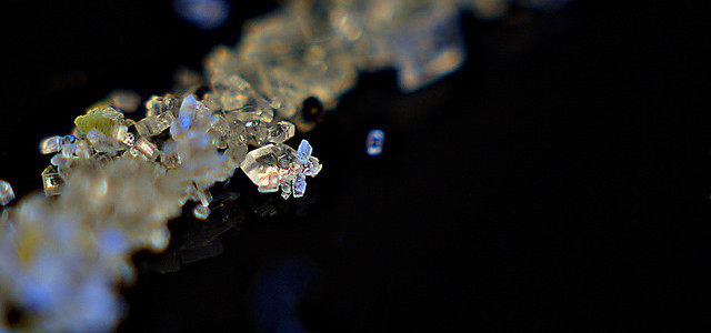
You do a Western blot looking at a new protein and instead of one specific band you see a few bands. One is close to the predicted molecular weight and is pretty distinct, but they others run more slowly indicating a greater molecular weight. The higher molecular weight bands are also kind of fuzzy.
Photo courtesy of Michael Pardo.
If you see this type of banding pattern, then you might suspect that your protein is glycosylated.
What is glycosylation?
Glycosylation is very common and occurs when cellular proteins covalently add sugar molecules onto proteins. The added sugars can be quite simple, or they can be complex with large bulky branches. The majority of proteins made in the rough endoplasmic reticulum (ER) are glycosylated.
What is glycosylation used for?
Glycosylation is believed to serve several functions:
- Aids in protein folding and stability
- Is a checkpoint for mis-folded proteins
- Alters protein:protein interactions
- Mediates cell:cell adhesion
- Mediates immune responses
- Alters solubility of protein
Several rare, congenital diseases are associated with improper glycosylation.
There are 5 types of glycosylation
There are 5 main types of glycosylation:
- N-linked: sugars are attached to the amino group of an asparagine (N) side chain
- O-linked: a monosaccharide is attached to the hydroxyl group of serine or threonine posttranslationally
- Glypiation: a glycosylphosphatidylinositol (GPI) anchor is covalently attached to a protein
- C-linked: a mannose sugar is added to a tryptophan through linkage to a carbon atom
- Phosphoglycosylation: oligosaccharides are linked to serine or threonine via phosphodiesters
Here were are just focusing on the two most common: N-linked and O-linked.
You gotta be exposed and in the right place
Glycosylation sites must be accessible to the glycosyltransferases and glycosidases for glycosylation to occur. Transmembrane proteins and secreted proteins are good candidates – they are made in the rough ER and portions of the proteins are exposed to the lumen of the ER and Golgi where the enzymes reside. As proteins fold though, potential glycosylation sites can become obscured or inaccessible.
Glycosylation is diverse by not random
Glycosylation does not occur randomly on a protein but is directed by certain consensus sequences. N-linked glycans occur on arginine residues contained in the following sequence: Asn-X-Ser/Thr/Cys; where X is any amino acid except proline. Although consensus sites are good predictors of glycosylation, not all of them are utilized. O-linked glycosylation doesn’t require a consensus site, but may occur on serines and threonines, and sometimes oxidized forms of proline and lysine.
The final composition of the glycan is diverse, particularly in N-linked glycosylation. Once the initial glycan has been attached, trimming enzymes (glycosidases) and other enzymes (glycosyltransferases) add and remove various sugars from the protein as it transits the secretory pathway. The final glycan composition is dependent on the enzymes involved, which can vary between tissue and cell types.
Stay tuned for our next article in which we will talk about different ways to study protein glycosylation.
Leave a Reply