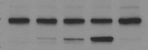
The Western blot is a useful technique for analyzing protein size and quantity, as well as assessing post-translational modifications. However, sometimes it is difficult to interpret your results because of high uniform background or uneven, splotchy background. In other posts we have given tips on how to avoid these common problems and we have also discussed this in detail in our October Wiki content.
A lot of these problems can also be avoided by having a standard protocol that can be tweaked for individual antigen:antibody combinations. Below is our recommended general protocol for Western blots, beginning with blocking and ending with detection.
1) Remove membrane from transfer set-up and place into clean incubation tray containing blocking buffer.
a. The appropriate blocking buffer should be determined for each antigen:antibody combination.
b. A general protein-based blocking buffer is 0.5-1% non-fat dry milk in phosphate buffered saline containing 0.05% Tween-20 (PBST)
2) Rock blot at room temperature for 1-1.5 hours.
3) Remove blocking buffer and rinse blot twice with PBST.
4) Incubate blot with primary antibody diluted in PBST for 1 hour at room temperature with agitation.
a. Primary antibody should be titrated for optimum concentration.
b. Make sure antibody solution completely covers membrane.
5) Wash blot 4 times with PBST; incubate 5 minutes at room temperature with agitation for each wash.
6) Incubate blot with secondary antibody diluted in PBST for 1 hour at room temperature with agitation.
7) Wash blot 4 times with PBST as described above.
8) During last wash prepare detection reagent according to manufacturer’s instructions.
9) Remove blot from wash and blot off excess wash.
10) Place blot into clean tray and add detection reagent (0.1ml/cm2 is usually sufficient to cover blot).
11) Incubate as directed.
12) Remove blot from detection reagent and blot excess reagent off membrane with filter paper. Wrap blot in plastic wrap.
a. Make sure the membrane does not dry out during manipulations.
13) Expose blot according to protocol.

Leave a Reply