Multi-color fluorescent Western blotting in one kit
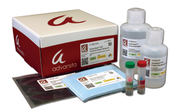
Try multicolor fluorescent western blotting today. Our kit includes everything except primary antibodies.
- Multiplexing – visualize two proteins simultaneously!
- Sensitivity –10x greater sensitivity than Cy dyes; pictogram detection
- Quality Reagents – includes AdvanBlock™-PF blocking and AdvanWash /AdvanWash-IR for washing
- Fast results – 3.5 hours for entire protocol
- Compatible with imaging systems that detect Cy3 and Cy5 (MCF) or near-IR imaging systems (MCF-IR)
- Fluorescent Detection – choose between visible (APC and RPE) or near IR (IR700 and IR800) kits with SpectraDye™ secondary antibodies
Kit Includes:
Secondary antibodies
Blocking solution
Washing solution
Pre-cut low-autofluorescence PVDF membranes
Background quenching sheets
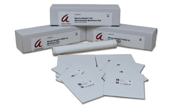
Convenient nitrocellulose and PVDF membranes for Western blotting
- Performance – quality controlled for background-free Western blots
- Convenience – membranes available pre-cut and as rolls
- High signal-to-noise ratio – low background signal for clearer results
- Flexibility – choose the right membrane for your Western blotting application
Membrane Options:
WesternBright PVDF-FL: fluorescent detection
WesternBright PVDF-CL: chemiluminescent detection; best stability for stripping and re-probing chemiluminescent blots
PVDF membranes available as rolls or pre-cut (7x9 cm, 10x15 cm, or 13x18 cm)
WesternBright NC: all-purpose nitrocellulose membrane
Nitrocellulose membranes available as rolls or pre-cut (8x10 cm)
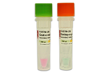
Fluorescently labeled secondary antibodies
- Fluorescent detection –secondary antibodies with a range of fluorescent conjugates
- Performance – strong signal and no cross-reactivity
- Convenience – each antibody demonstrated to provide optimal results with Advansta's Western blotting systems
- Flexibility – choose from visible or near IR fluorescent dyes and target species best suited to your application (see product listings)

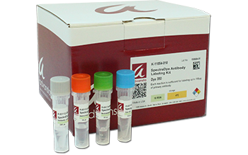
Make your own fluorescent antibody in one easy step.
- Quick and easy – one-step labeling in 30 minutes
- Versatile - Labeled antibodies are compatible with immunofluorescence microscopy, flow cytometry, Western blotting
- Flexible - choose from 6 commonly used fluorescent dyes
- Save time and money – no secondary or extra steps, once you label your primary
- Save antibody – labeling reaction requires as little as 10 µg antibody
Kit includes:
Antibody Labeling Buffer
SpectraDye Dye Solution*
Quenching Solution
Neutralization Buffer
Sufficient reagents to label up to 1 mg of antibody
*Except kit K-11060-010
LightSaver™ Fluorescence Enhancing Solution
- Enhances – fluorescent signal
- Compatible – with PVDF and Nitrocellulose membranes
- Convenient – ready-to-use solution
- Compatible – with visible and near IR fluorescent westerns
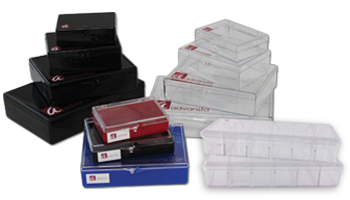
Convenient containers for staining and washing gels and membranes
- Smooth interior – protects membranes and gels from scratches
- Convenient – multiple sizes (5x7, 6x9, 9x11,10.5x15.5 cm) and designs available for different gels and blots
- Attached lid – to protect experiments from dust or debris that can cause speckles on blot images
- Save – minimize antibody and buffer usage by using appropriately sized containers
- Multi-chamber trays – process 6 mini-blots simultaneously, each chamber requires as little as 3 mL of solution
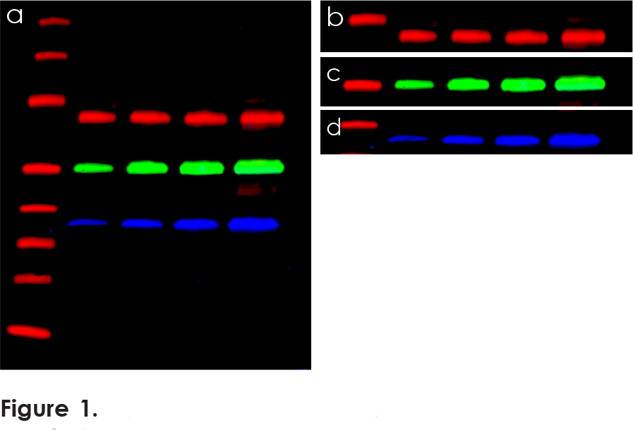
Ready-to-image three-color fluorescent blot
- Fluorescently labeled primary and secondary antibodies fluoresce in the red, green, and blue fluorescent channels (like Cy2, Cy3, and Cy5 equivalent dyes)
- Convenient – an easy way to evaluate the performance of your fluorescence imaging system
- Reliable – quality controlled, with lot-specific statistics provided for every blot
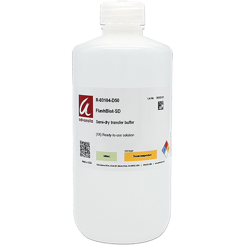
Rapid high efficiency semi-dry transfer buffer
- Fast - high ionic strength formulation allows for protein transfer in 3 to 10 minutes when used with a compatible high current semi-dry system
- Compatible - use your existing high current semi-dry transfer apparatus
- Reproducible - consistent transfer across entire blot
- Versatile - use nitrocellulose or PVDF membranes to achieve transfers with low background and high sensitivity with both chemiluminescent and fluorescent Western blots
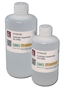
Ready-to-use wet transfer buffer
Designed for faster, more efficient wet protein transfer with increased sensitivity
FLASHBlot is not your average wet transfer buffer.
- Faster than traditional wet buffers: better results in 20 minutes
- More sensitive detection for low abundance proteins
- Better detection of post-translationally modified proteins
- Better transfer of ALL proteins, including high molecular weight proteins
- Conveniently suitable for any wet transfer set-up
“ We were pleasantly surprised to see that with FLASHBlot we observed the same transferring levels at 25 min than with the lengthier transfer protocol… including for the 300 kDa protein. This reagent will shorten considerably our experimental time. ” - Assistant Professor, UCSF
Transparent plastic supports for handling and imaging blots
- Clear images – avoid the wrinkling or bunching that can happen with plastic wrap
- Suitable for all blots – development folders are suitable for chemiluminescence and fluorescence imaging
- Safe blot handling – move your blot without scratching or putting pressure on the surface
- Convenient size – image up to 2 blots simultaneously in one folder
Pre-cut paper for electrophoretic transfers
- Convenient – pre-cut; work with most mini gel transfer assemblies
- Standardized – uniform thickness for reproducible transfer
- Tested – perform consistently without artifacts or background
- Available sizes include 7x9 cm and 8x10 cm
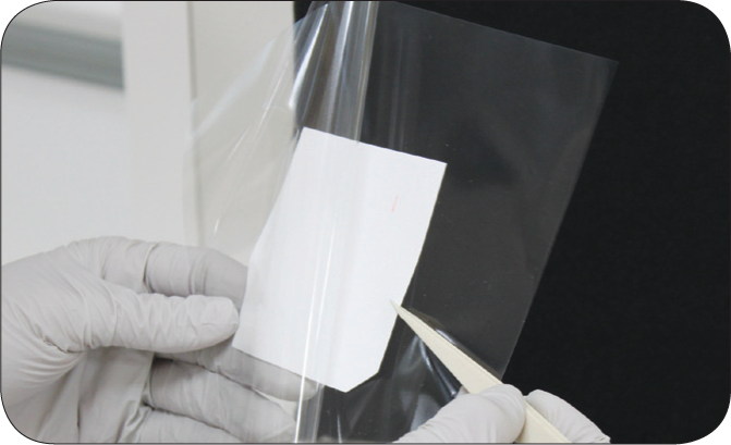
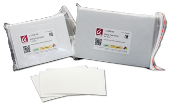
An added layer to decrease background
For improved imaging of blots and gels
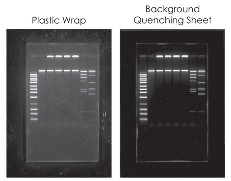
- Better images by removing background fluorescence and stray light
- Improved signal to noise ratios
- Simple – just place sheet under the blot or gel
- Compatible with chemiluminescent and fluorescent Western blots
- Compatible with DNA gels and protein stains - including SYBR green, SYBR Gold, ethidium bromide, SYPRO® Orange, SYPRO Ruby and more



Connect